
의학/case review
diverticulitis 22/07/02
Hong :)
2022. 7. 2. 18:59
728x90
반응형
CASE1 >
F/39
응급실 기록
내원 당일 오후 6시 부터 시작된 flank pain을 주소로 본원 응급실 내원함.
RLQ pain 동반되며 속이 메스꺼워 어제부터 식사를 잘 하지 못했다고 함.
BT 38.2
P/Ex RLQ Td (+) N/V (-/-) Urinary symptom - c/s/r -/-/-
응급실 검사
CRP (C-Reactive Protein), quan. 77.81 ▲
WBC Count 11.29 ▲
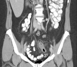
APCT >

Adm 기록 GS
입원하여 conservative care
게실에 대한 평가 위하여 증상 회복 후 대장내시경 f/u이 필요함을 설명함.
-> 여러 번 재발시 장 절제 수술
CASE2>
Case review > diverticulitis
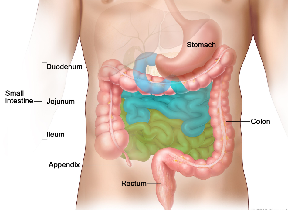
diverticulosis : diverticula가 있는것
diverticulitis : 염증이 생긴 것
- Sx : abd pain, Td, fever, leukocytosis / 진행될수록 symp more generalized, widespread
What are the imaging studies to detect findings of diverticulosis?
- Lower GI EGD
- CT
What are the imaging findings of diverticulitis?
- US: Abnormal wall thickening of more than 4 mm involving a segment 5 cm or longer at the point of maximal tenderness.
- CT:
- Diverticula
- Narrowed lumen
- Segmental Thickened bowel wall : usually has inner and outer high-attenuation layers, with a thick middle layer of low attenuation
- Fascial inflammatory infiltration
- Complications
- Perforation: Free air in the peritoneum
- Abscess
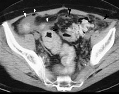
Perforated Diverticulum
- Arrowheads point to free air.
- Arrows points to collection of fluid around bowel loops.
- Black arrows point to pericolonic fascial infiltration.
728x90
반응형